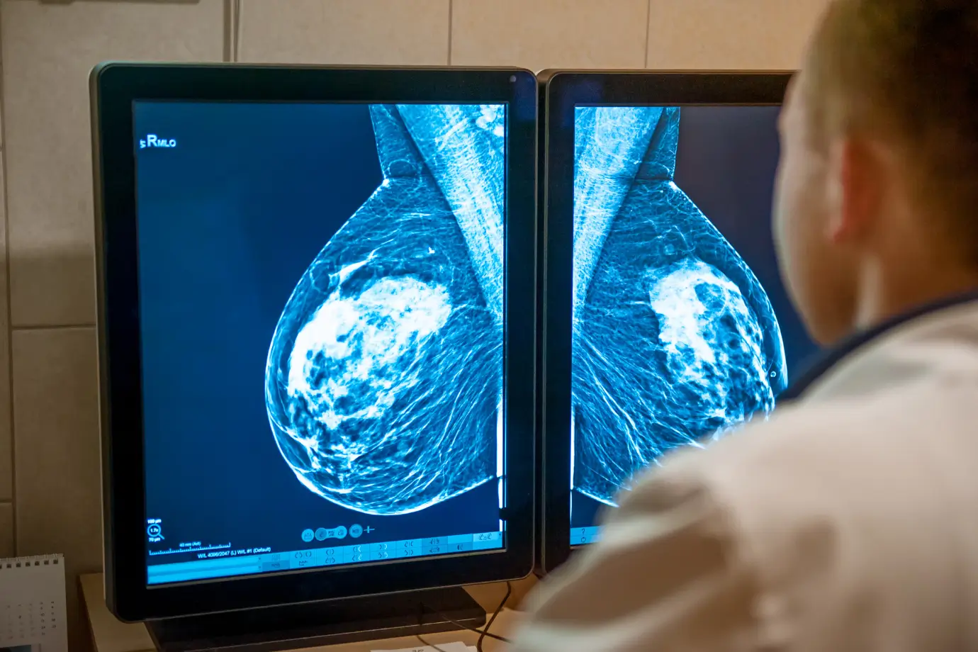
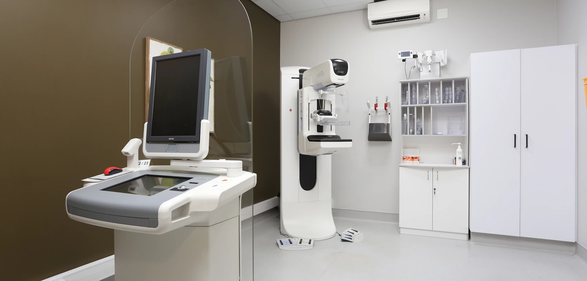
Radiology Services

Mossel Bay Radiology Services

Advanced radiology services with a patient-first approach.

Mammography

Mammography is a well-established screening and diagnostic tool used to evaluate breast tissue for the possible presence of cancer through the use of low-dose X-rays.

The goal of mammography is to aid in the early detection of breast changes and doing so, improve prognosis.

We utilize tomosynthesis with our mammograms, which is an advanced form of mammography providing thin slice evaluation, improving detection rates, and reducing the need to perform additional mammographic views.

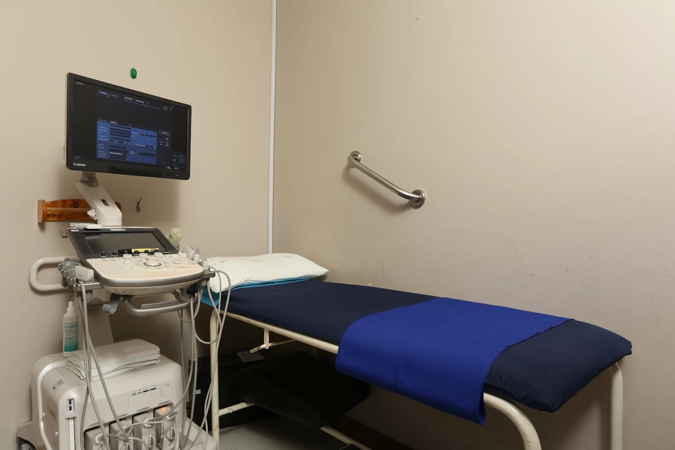
All our mammograms are followed by an ultrasound examination to allow for further analysis and, if necessary, lesion characterization.

All mammograms and ultrasounds are performed by trained mammographers and reviewed by a radiologist.

We follow international guidelines and recommend an annual mammogram from age 40. Rarely, in patients at higher risk, earlier screening mammograms might be indicated.

If a lesion is detected, we offer imaging-guided biopsies, including ultrasound-guided and stereotactic (mammogram) guided biopsies.

If your last mammogram was performed elsewhere, please bring the CD or films along, if available.

Do not wear deodorant, lotion or use talcum powder on the day of examination, as they can cause artefacts, limiting evaluation.

You should inform the mammographer if there is any chance you might be pregnant.

Mammography

Bone Densitometry

It is performed to assess for osteoporosis or to monitor the treatment thereof.

It uses two low-dose X-rays of different wavelengths to measure the calcium content of certain areas of the body - typically the lumbar spine, hips or wrists.

It is a quick and painless examination.

Patients should ideally not have the study performed within 10 days of having any barium examination or contrasted CT.

Bone Densitometry

CT Scan

A CT scan uses X-ray technology to produce cross-sectional images of the body part being imaged, while the patient moves through the gantry on the examination bed. Allowing for analysis of the organs, soft tissues and the skeleton/bones.

Often, intravenous contrast is administered on the referring doctor's request, which allows for further and more detailed analysis.

As per international guidelines, certain patients might be requested to do a blood test prior to the examination if there has not been a recent test, to check their kidney functions, as the intravenous contrast is cleared from the body by the kidneys mostly.

The intravenous contrast can cause a warm feeling throughout the body and a metallic taste in the mouth, which is normal and usually clears within a short while.

Please inform the radiographer if there is any chance you might be pregnant.

CT Scan

Fluoroscopy

Fluoroscopy uses low-dose X-rays to obtain real-time/live images of the body part being examined, similar to a movie. It is often used to assess swallowing and the function of the oesophagus, or to assess certain bladder functions, however, it has many applications.

Patients are often given dilute Barium (which is a naturally occurring element that shows up well on X-rays) to swallow during the examination. Other contrast agents can be used depending on the examination requested.

Fluoroscopy

X-rays

Radiographs are a pain-free examination using X-rays to image the body region in question - for eg joints, chest, and abdomen.

The examination involves exposing the body part in question to a small dose of ionizing radiation to obtain the image. The amount of radiation is negligible, and we adhere to local and international guidelines and protocols.

Please inform the radiographer if there is any chance you might be pregnant.

X-rays

Interventional Radiology

Interventional radiology is a term encompassing multiple minimally invasive procedures - extending from imaging-guided biopsies of lesions to drain placement and imaging-guided lumbar punctures.

These are performed upon the request of the referring doctor.

Please inform the radiographer if you are at an increased risk of bleeding or if there is a chance you might be pregnant.

Interventional Radiology

MRI

MRI or magnetic resonance imaging uses electromagnetic fields and radio waves to obtain images. It does not use ionizing radiation.

It is used for many body regions, including the head, spine, liver, heart, joints and breasts.

In addition to anatomical information, it also provides certain functional/physiological information.

Occasionally, intravenous gadolinium might be administered to provide further relevant information, depending on your doctor's request.

Because of the magnetic fields, certain types of pacemakers, cochlea implants or patients with shrapnel, for example, might not be able to undergo an MRI examination - for the patient's safety. This can be discussed at the time of booking, and if needed, alternative imaging recommended.

It is particularly important with MRI that the patient move as little as possible during the examination, as this causes significant artefacts.

Patients should please not wear mascara on the day if booked for examination of the head or neck, as this can cause artefacts.

If you suffer from claustrophobia and might request light sedation, please bring someone with you to drive you home, as you may not drive for 24 hours post-sedation.

You will be provided with a panic button should you need immediate assistance from the radiographer.

The machine is similar in appearance to the CT scanner, with a donut shape, the centre of which the bed/patient moves through. It is open on both ends.

Sometimes, when a joint is being imaged, it might be necessary to inject fluid into the joint, called an arthrogram, to aid in further evaluation.

MRI

Ultrasound

Ultrasound imaging utilizes sound waves for a safe, painless examination that assists in the diagnosis and follow-up of many conditions.

It does not use ionizing radiation and is therefore safe in pregnancy.

If you are booked for an abdominal ultrasound, if possible, do not eat for 6 hours prior to the examination so that the stomach is not full. If booked for abdominal/pelvic or kidney ultrasound, please drink 4-6 glasses of water in the hour before the examination so that your bladder is full.

Ultrasound examinations are performed by qualified sonographers or radiologists.

Ultrasound